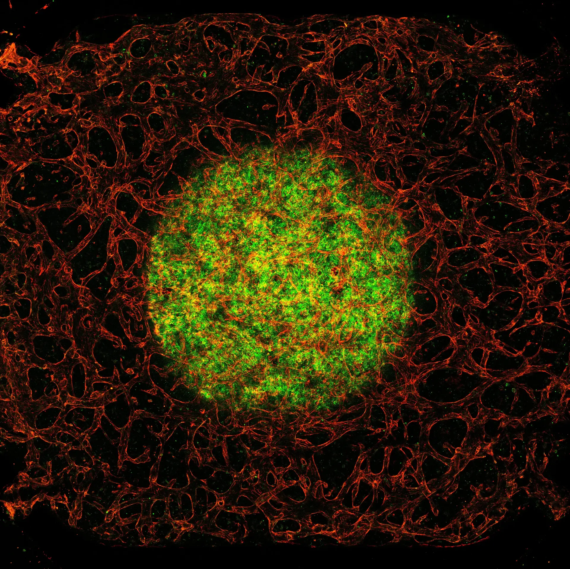
Fluorescence microscopy image by Laurens Spoelstra
Laurens Spoelstra  ·  Zeiss LSM880
Fluorescence microscopy image by Lena Koch
Lena Koch  ·  Zeiss LSM880
Fluorescence microscopy image by Lena Koch
Lena Koch  ·  Zeiss LSM880
Fluorescence microscopy image by Tarek Gensheimer
Tarek Gensheimer  ·  Zeiss LSM880
Fluorescence microscopy image by Tom Luiken
Tom Luiken  ·  Zeiss LSM880
ML-I FACILITY

BioImaging
Centre

State-of-the-art light microscopy, experimental imaging techniques, image analysis and cell/tissue culture facilities.

15+
Advanced Imaging Systems
24/7
Access for Trained Users
NL-BioImaging
Affiliated Infrastructure
UT + External
Users Welcome
Welcome

A shared facility for cutting-edge imaging

The BioImaging Centre (BIC) is a Multi-Level I facility offering state-of-the-art commercial microscopes, custom experimental setups, advanced image analysis, and ML-II cell & tissue culture infrastructure.

Open to both University of Twente researchers and external users. Watch the video to get a feel for our facility and what we can offer your research.

How to get started
Analis
Official Analis Experience Center
Unique in the Netherlands
Why Choose BIC

Capabilities you won't find anywhere else in the Netherlands

From specialised optics to a complete end-to-end research pipeline — BIC offers unique infrastructure designed to take your science further.

Unique in NL

Wavefront Shaping in Two-Photon Microscopy

Our two-photon microscope is equipped with adaptive wavefront shaping — a powerful technique that corrects optical aberrations to achieve diffraction-limited imaging deep into scattering tissue, a capability unavailable in standard facilities.

Specialised Optics

High-NA Deep-Tissue Objective

We house the Evident XLSLPLN25XGMP — a 25× objective with NA 1.0 and an exceptional 8 mm working distance. This rare combination delivers high-resolution imaging deep into cleared tissue or brain samples, without the usual trade-off between resolution and penetration depth. Paired with a custom water bridge from Life Imaging Services, making it fully practical for routine deep-tissue experiments.

End-to-End

Complete Research Workflow

BIC provides the full pipeline under one roof — from cell culture and sample preparation in our ML-II facility, through imaging on state-of-the-art microscopes, to quantitative image analysis. No need to move between institutions.

Portfolio

Our Equipment

Every system in the facility, grouped by technique — with the specifications, typical applications and access notes for each. If you are not sure what your experiment needs, describe it and the Microscope Finder will suggest a match.

Grouped by technique
  • Widefield & Brightfield
  • Confocal & Two-Photon
  • Specialized & Experimental
  • Flow Cytometry

Biological Sample Facilities

ML-I Facilities

  • • Imaging inside Laminar Flow Hoods (Echo Rebel)
  • • Balances, pH-meters, pipet sets
  • • Fume Hood for chemical sample handling
  • • Basic consumables (tips, gloves, slides)

ML-II Facilities

  • • CO₂ Incubators
  • • Laminar Flow Hoods
  • • Centrifuges & Freezers
  • • CRISPR-Cas9 available on request

Image Analysis

The BIC provides Imaris Essentials 9.9.1 image analysis software free of charge to all users. The software can be used on the dedicated Imaris PC in ZH161 or via Remote Desktop.

We also support ImageJ, FIJI and MicroManager. Our technicians provide training and help with experimental design and custom protocols.

Pricing

Rates

The BIC is open to both University of Twente researchers and external users. Rates depend on your user type and the equipment used — contact us for a tailored quote.

Internal
UT Researchers
  • Preferential rates for University of Twente staff and students
  • Access to all microscopes after introduction & training
  • Free Imaris image analysis software
  • Technical support from BIC staff
  • ML-II cell culture facilities available
External
Industry & Other Institutions
  • Open to companies, hospitals, and other research institutes
  • Full access to the BIC equipment portfolio
  • Imaris image analysis available on-site
  • Expert guidance and assisted sessions available
  • Custom imaging projects & collaborations welcome
Contact us for rates

We'll get back to you with pricing tailored to your user type and needs.

How to use the BioImaging Centre

New to the BIC? Start by exploring our equipment to find the systems that fit your research. Once you know what you need, fill in the entry form below and we'll contact you to schedule an introduction and training.

Open Entry Form
Opening hours

Monday – Friday 08:00 – 18:00
Extended access possible for experienced users.

Rates

Internal and external rates available on request. See pricing →

LEARNING CENTER

Understand it before you book it

Interactive simulators that let you change one thing at a time and watch what it does to your image. Start with the Coverslip Focus Lab and see why the glass you choose costs you more signal than you would guess.

Research Impact

Featured Publications

Highlighting the great science happening with BIC — your work could be next.

Used our facility in your research? We'd love to be acknowledgedget our suggested citation.

Advanced Healthcare Materials · 2026

A Synovium-on-Chip Platform to Study Multicellular Interactions in Arthritis

Spoelstra LR, Semmekrot FR, Araújo-Gomes N et al.

DOI: 10.1002/adhm.202504857 Read paper
"Stained chips were imaged at the University of Twente BioImaging Centre facility using a Zeiss LSM880 laser scanning confocal microscope."
Lab on a Chip · 2026

Label-free assessment of a microfluidic vessel-on-chip model with visible-light optical tomography reveals structural changes in vascular networks

"Microscopy work for this study was supported by the BIC at the University of Twente."

Veerman D, Cuartas-Vélez C, Gensheimer T, van Dorp T, van der Meer A, Bosschaart N

DOI: 10.1039/d5lc00927h Read paper
Advanced Healthcare Materials · 2026

A Quantitative Printability Framework for Programmable Assembly of Pre-Vascular Patterns via Laser-Induced Forward Transfer

"Part of the microscopy work for this study was supported by the BIC at the University of Twente."

Bosmans C, Ginés Rodriguez N, Gutiérrez Hernández UJ, Fernandez Rivas D, Karperien M, Malda J, Moreira Teixeira L, Levato R, Leijten J

DOI: 10.1002/adhm.202503665 Read paper

Partners & Collaborations

Analis
Analis
Experience Center
NL-BioImaging
NL-BioImaging
Affiliated Infrastructure

Get in touch

Questions about your imaging project or want to discuss possibilities?

TNW-BIC-Support@utwente.nl Follow us on LinkedIn
053 489 1262
053 489 3021
Zuidhorst ZH161, De Horst 2, 7522 LW Enschede  ·  Parking P4

How to get here

1
Arriving at the campus

Follow the signs for "Other buildings" until you cannot go further.

2
Parking Lot 4 (P4)

You'll reach Building 28 (ZuidHorst) on your right, with P4 directly next to it.

3
Find us inside

The BioImaging Centre is inside ZuidHorst, room ZH161.